Reports of a chilblain-like illness in young people and children linked to the coronavirus pandemic.
COVID-19 continues to spread across the globe, infecting people on every corner of the planet. The general perception is that the disease is less severe and often mild in younger patients. Whilst this is usually the case, it does not mean that younger patients always escape from complications. One potential COVID-19 complication, which is being reported with increasing frequency in children and young adults, is “COVID toes”. A new study published in the British Journal of Dermatology takes a deeper look at this phenomenon and how exactly viral infection leads to chilblain-like lesions on the toes (1).
“COVID toes” is the term commonly being used to describe inflammatory lesions seen on the toes of many young people during the COVID-19 pandemic. Most of these patients do not suffer any respiratory symptoms and in many cases, swabs taken from the nose or respiratory tract of these patients test negative for SARS-CoV-2. This has given rise to a number of important questions. Foremost amongst these are the questions of whether COVID toes phenomenon is actually caused by SARS-CoV-2 and if so, how is it that respiratory samples from these patients regularly test negative?
To answer these questions, a research team retrospectively examined the details of all cases of pediatric chilblains diagnosed at a hospital in Madrid over a four-week period from April to early May 2020. This included a total of seven pediatric patients ranging from 11 to 17 years of age. In all cases, the lesions were not particularly painful or itchy and resolved over time without any harmful outcome to the patient.
Nasal and oropharyngeal swabs were tested in six of the seven cases and in all six cases, the samples tested negative for SARS-Cov-2. However, these patients also had biopsy samples taken from the chilblain lesions. Cellular material from these biopsy samples tested positive for the presence of SARS-CoV-2 elements, most notably the “spike protein” the virus uses to attach to and enter human cells. Further examinations of the biopsy material revealed that the inflammation was caused by the accumulation of inflammatory T cells and lymphocytes.
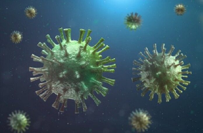
Using an electron microscope, they also examined the cytoplasm of cells from the lymph vessels in the chilblain regions. This examination revealed the presence of small, spikey particles that match both the size and shape of the SARS-Cov-2 virus.
The evidence uncovered in this study suggests that although patients may test negative for SARS-CoV-2 using the routine nasal or oropharyngeal swabs, they may still have been infected. The chilblain lesions themselves were the result of inflammation of lymph vessels, likely caused by the presence of the virus. Chilblains are usually classified as primary (no identified cause or caused by cold temperatures) or secondary (where an underlying cause has been identified. Secondary causes include inflammatory conditions such as lupus erythematosus or rheumatoid arthritis. However, none of the seven patients in this study had a pre-existing diagnosis of such conditions.
The absence of any causative condition, coupled with the detection of SARS-Cov-2 protein in biopsy samples strongly supports the authors’ contention that these lesions are in fact caused by the coronavirus. However, interpretation of the results must be done cautiously. The sample size in this study (n=7) was small and came from a single population at a single hospital. Further research will be needed to confirm these results and to shed more light on why children and young adults appear to be affected by this manifestation of SARS-CoV-2 whilst escaping the more harmful respiratory effects.
Written by Michael McCarthy
1. Colmenero I, Santonja C, Alonso-Riaño M, Noguera-Morel L, Hernández-Martín A, Andina D, et al. SARS-CoV-2 endothelial infection causes COVID-19 chilblains: histopathological, immunohistochemical and ultraestructural study of 7 paediatric cases. British Journal of Dermatology.n/a(n/a).
Image by Monoar Rahman Rony from Pixabay


