A non-invasive diagnostic test for endometriosis could soon be on the way. Earlier this year, Oxford University researchers presented preliminary data from a clinical trial supported by Serac Healthcare. Researchers hope that this scan based diagnostic will provide faster answers for women living with this painful condition. Dr. Druin Burch Chief Scientific Officer of Serac Healthcare took some time to tell us the story behind the science.
Around 10% of women of reproductive age develop painful growths of tissue similar to the lining of the uterus (endometrium), seemingly at random in their abdominal cavity and pelvis. What causes it and how it develops is still a mystery. The most common symptoms include abdominal and pelvic pain that comes and goes and difficulty conceiving.
Right now the only way to diagnose all stages of the condition is laparoscopy. This is a surgery performed under general anaesthetic. If doctors need to rely on patients having an operation to even know if they have a disease, it presents a huge problem. So why is surgery the only option? In short they are working blind.
A Painful Condition
Endometriosis, in simple terms, is when tissue that looks similar to the lining of the womb, grows outside the uterus. Lesions can form on your intestines, your bladder, the exterior of your uterus, basically anywhere in your abdomen or pelvis. This tissue shares many features of endometrium – including the growth of new blood vessels. This means sex hormones like oestrogen and progesterone circulating in your body, trigger the endometriosis patches to build themselves up and then shed. The build and shed create painful lesions in your abdomen, inflammation and scarring where the body is trying to heal itself.
On average, it takes a patient seven and a half years to be diagnosed with the disease. In conversation with MNB, Burch explained that this is down to lack of education about endometriosis and the technical challenges of making a diagnosis. Endometriosis symptoms are vague and vary so much between individuals that it’s not usually the first ailment family doctors will consider. Endometriosis is hard to spot, the symptoms overlap with so many other conditions, and the only way to definitively diagnose it is keyhole surgery. This is a procedure performed under general anaesthetic. A surgeon will make incisions into the patient’s abdomen and insert a camera so the team can directly look at internal organs. If they spot scarring or active lesions, you will get a diagnosis of endometriosis. If there is no sign of scarring or lesions, you will not be diagnosed with endometriosis.
Blind Guesses
Since the only way to identify all stages of endometriosis including early disease is keyhole surgery, getting tested is an ordeal. It’s expensive, there are always risks to undergoing surgery, and elective surgical procedures often have long waiting times. What’s more, Burch explained, up to 50% of women referred for a laparoscopy don’t have any visible lesions. This means a whole lot of waiting, having surgery and recovery to be told their symptoms were not due to endometriosis after all.
Not only is endometriosis hard to diagnose, it’s incredibly difficult to research. If the only way to watch it progress, take tissue samples or even see if someone has it, is a laparoscopy, how can researchers ethically study it?
Imaging specialists at Serac Healthcare in collaboration with University of Oxford are on their way to solving this problem. They have developed a way to label potential endometriosis that will allow doctors to use a body scan similar to a CT scan to spot lesions. What’s special about this approach is that they will use a brand new molecular label to highlight tissue that behaves like endometrium. This imaging agent has never been used before in endometriosis detection and will be the first imaging label to allow medics to accurately see endometriosis without surgery. The researchers at Serac hope that this will lead to easy and fast diagnosis of this tricky to identify disease. They also intend to give researchers a tool to learn about the condition in ways that have never before been possible.
X-ray Vision
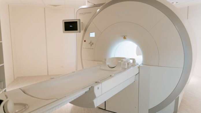
Using their brand new molecular imaging label, the team at Serac Healthcare plan to employ widely available medical diagnostic imaging equipment that is already in many hospitals. Using a SPECT/CT scanner, the doctors will hunt down patches of endometrium-like tissue with the aid of a special molecule that labels newly made blood vessels. A patient will be injected with a radio-labelled imaging agent and then hop into an imaging machine that will scan their body taking digital photographs. A software program then builds 3D reconstructions of all the places in the body indicating where the agent was detected. A radiologist will examine the images for bright spots. These bright spots should correspond to patches of new blood vessels. The doctors will then make a judgment call on whether it looks like an endometrial lesion.
Maraciclatide – A Long Time Coming
The most important element of the test is the labelling molecule – Maraciclatide. Maraciclatide works by binding to newly formed blood vessels (a process called angiogenesis). It was originally developed as a potential cancer detecting tool, but didn’t find its niche. Dr. Burch recounted how, some of the researchers who worked on Maraciclatide saw its potential beyond oncology.
“[They] felt it had tremendous potential in a number of conditions,” he says, “so they acquired the rights to develop the molecule themselves.” He went on, “They were so committed to the idea that there were major medical opportunities here, that they quit their jobs to develop it themselves.”
Initially the researchers planned to use Maraciclatide, a short peptide that binds to a protein found in tissues in which new blood vessels are developing, to look at rheumatoid arthritis. Rheumatoid arthritis is a form of chronic inflammation. Chronic inflammation can trigger angiogenesis – the formation of new blood vessels. In turn angiogenesis can trigger inflammation, this creates a feedback loop that amplifies the inflammation. The inflammation triggered when new blood vessels form, is a feature shared by endometriosis. As Burch explained, “It was a natural progression. When we got the molecule it was already slightly more advanced in terms of its development for inflammatory arthritis, and it has remained that way, but endometriosis had been identified at a very early stage of the molecule as a setting in which it might be useful.”
R&D
The scientists soon made moves to investigate this multitasking molecule’s potential for endometriosis imaging. This was not an easy process, however. “From the moment of the company being set up there was always active interest in trying to work out whether we could be helpful in endometriosis. But it takes a while to raise money and get studies under way.”
The team first had to find investors willing to take a risk on this common but under studied disease. As Burch explained, “Raising money for a biotech start up is always a challenge. Yet the management team successfully raised the necessary funds for the initial research and development in inflammatory arthritis and endometriosis. There is also the wider issue of historical underfunding of endometriosis research, however, clearly, there’s huge enthusiasm at the moment to change that, to try and inject more interest and more funds into researching endometriosis. And certainly, a lot of our investors have been really enthusiastic in seeing if we can make a difference in that area.”
Next, Burch got to work tracking down assistance from medical researchers ready to do independent clinical trials to put the test into practice.
Serendipity
By happy accident, both Burch and the Oxford Endometriosis CaRe Centre, a
world-leading endometriosis group are based in Oxford, United Kingdom. Serac Healthcare connected with the team recruiting them to test out their imaging technique. Since Maraciclatide has already been investigated as an imaging agent for cancer and inflammatory arthritis, the researchers at Serac and University of Oxford have hit the ground running on endometriosis. With extensive safety data from various clinical trials and phase III clinical trials about to start in inflammatory arthritis, the researchers could get straight into figuring out whether this approach works on endometriosis.
As we reported in April this year, Dr. Tatjana Gibbons working with Professor Krina Zondervan and Professor Christian Becker, Co-Directors of the Oxford Endometriosis CaRe Centre, presented encouraging results at the International Society for Reproductive Imaging meeting.
The Oxford researchers are currently conducting the DETECT study (Detecting Endometriosis expressed integrins using technetium-99m). Burch told us, “[they] are aiming to recruit up to about 25 volunteers who are going to be having a laparoscopy anyway, and asking them if they’d be willing to have a scan just prior to their laparoscopy, and then comparing the results to the surgery and seeing if we can see disease in the same places”.
But reality is never so simple, this is an ambitious project. There are currently no established imaging parameters for endometriosis. Before this test can progress to phase III clinical trials, the researchers must find out how best to use the imaging agent: technical wrinkles must be ironed out. For example, how much imaging agent do researchers need in order to see anything, how long should they wait before jumping into the SPECT/CT scanner for imaging? Despite these challenges it seems that this test is making progress.
Encouraging Results
Dr. Gibbons presented preliminary data indicating that they have jumped the basic hurdles and are currently able to detect superficial peritoneal lesions – an early form of endometriosis that currently can only be seen reliably during laparoscopic surgery. Zondervan and Becker anticipate that the trial will be complete by the end of the year.
While we might not be seeing phase III trial results for some time, the news from Zondervan and Becker’s research initiative is reason to celebrate.
Burch says, “the early results that we have are hugely encouraging and so we’re very optimistic. They are early results – however we are encouraged that it might be useful in a number of different settings. One setting would be the diagnosis of the condition, so hopefully reducing that seven to eight years’ delay, by addressing the unmet need for a non-invasive test for endometriosis.’’


