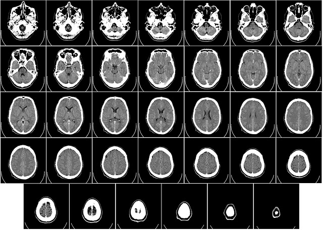
Advanced Magnetic Resonance Imaging (MRI) brain scan analysis helps to identify patients with a high risk of developing stroke-related dementia, study finds.
Strokes, which occur when blood flow to the brain is blocked, can affect the thinking abilities of the patient and also lead to dementia. When the stroke affects the small blood vessels in the brain it is known as cerebral small vessel disease. Atherosclerosis or plaque building up in the small blood vessels in the brain could result in the vessels being blocked and the brain starved of oxygen (ischemia), or the vessels leak causing bleeding or hemorrhage.
Cerebral small vessel disease is associated with 45% of dementia cases and about 20% of strokes worldwide. Patients show a decrease in cognitive abilities. Diagnosis of small vessel disease relies on imaging techniques such as an MRI scan. Markers associated with these disease abnormalities appear as imaging changes in different areas of the brain as observed in an MRI scan. Using information from these different changes to develop a single cumulative marker has the potential to improve diagnosis.
Researchers from the University of London, UK have assessed the use of one such imaging method known as diffusion tensor image segmentation (DSEG) technique to predict the risk of developing dementia and the level of cognitive decline in patients with cerebral small vessel disease. This MRI scanning method allows for the characterization of micro-structural features of the brain. The researchers used the information obtained from the scan to develop a single DSEG score that reflects changes in the different markers of the small vessel disease. The researchers note that the combined score is “a more accurate method for monitoring disease progression […], with better predictions of cognitive decline than any single magnetic resonance marker”. Evaluation of this technique with patients enrolled in the St George’s Cognition and Neuroimaging in Stroke (SCANS) study between the years 2007 to 2015 in London. The results of this study were published in the journal Stroke.
The study included 99 patients with small vessel disease caused by ischemic stroke, with an average age of 68. The study participants received MRI scans for three years and underwent cognitive testing for five years. Amongst these participants, eighteen patients developed dementia during the study period, with an average time to onset of a little over three years. The DSEG score combined with the cognitive testing revealed that the patients with the most brain damage and poorer cognitive abilities at the start of the study were at much higher risk of developing dementia. The analysis could predict three-quarters of the dementia cases that occurred over the duration of the study.
The reference for comparisons at the start of the study was the youngest patient with least amount of brain damage. The single point of reference and the small subset of patients included in this study could mean that the results may not apply to patients with different forms of this disease. However, the potential of the method to accurately predict the risk of developing dementia will help identify patients at risk early on and early treatment could slow the progress of mental decline in these patients. As the senior author of the study, Rebecca A. Charlton, Ph.D. noted, “We have developed a useful tool for monitoring patients at risk of developing dementia and could target those who need early treatment.”
Written by Bhavana Achary, Ph.D.
References:
Williams OA, Zeestraten EA, Benjamin P, Lambert C, Lawrence AJ, Mackinnon AD, Morris RG, Markus HS, Barrick TR, Charlton RA. Predicting Dementia in Cerebral Small Vessel Disease Using an Automatic Diffusion Tensor Image Segmentation Technique. Stroke. 2019 Sep 12:STROKEAHA119025843.
Press release retrieved from https://www.eurekalert.org/pub_releases/2019-09/aha-amb090919.php
Information on cerebral small vessel disease.
Information on the prevalence of cerebral small vessel disease in patients with dementia and strokes obtained from-
de Leeuw FE, de Groot JC, Achten E, Oudkerk M, Ramos LM, Heijboer R, Hofman A, Jolles J, van Gijn J, Breteler MM. Prevalence of cerebral white matter lesions in elderly people: a population based magnetic resonance imaging study. The Rotterdam Scan Study. J Neurol Neurosurg Psychiatry. 2001 Jan;70(1):9-14
Image by WikiImages from Pixabay


