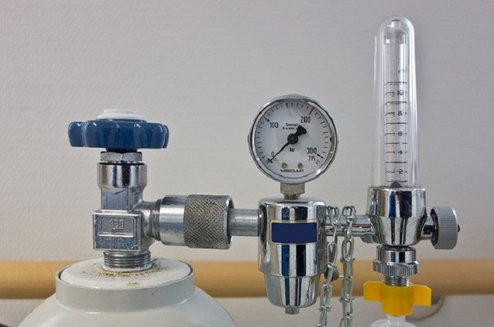
A ventilator is a life support machine that helps a person breathe when they are not able to do so on their own – how does it work?
Respiratory failure is a medical condition in which the respiratory system is unable to provide enough oxygen to the blood or eliminate carbon dioxide from the blood. The respiratory system comprises organs and tissues involved in breathing. These include the lungs, responsible for gas exchange between oxygen and carbon dioxide, and the pump – chest wall and abdominal muscles that help in the movement of air into and out of the lungs during breathing. Ventilators work to help with this process when the body cannot.
Lung failure, caused by diseases such as pneumonia, pulmonary (lung) scarring and emphysema, lead to impairing the gas exchange process. This results in dangerously low oxygen levels in the blood. Failure of the respiratory pump can result in insufficient ventilation of the lungs, leading to elevated levels of carbon dioxide in the blood. This is generally caused by medical conditions such as sleep apnea, hypothyroidism, injuries to lungs as well as the surrounding bones and muscles and blocking or narrowing of airways.
In some cases, individuals may experience respiratory failure in which their oxygen levels are too low and the carbon dioxide levels are too high at the same time. This can generally occur in patients with conditions such as asthma, chronic obstructive pulmonary disease (COPD) and pulmonary edema.
Respiratory failure is diagnosed by tests involving measuring the levels of oxygen and carbon dioxide in the blood as well as chest x-rays to determine the cause. Treatments for this condition include mechanical ventilation and providing supplemental oxygen. Mechanical ventilation is generally administered to patients placed in an intensive care unit (ICU) and may also be given to patients put under general anaesthesia during a surgery.
How does a ventilator work?
A mechanical ventilator is a medical device that assists in or takes over the task of breathing when the individual is unable to breathe on their own. The ventilator helps lower the amount of effort taken for the patient to breathe, allowing their body to recover from the illness or injury. It helps move air with increased levels of oxygen into their lungs, using positive pressure. It can also be used to remove carbon dioxide from the body.
The ventilator is connected to a monitor, which displays the patient’s vitals such as blood pressure, heart rate, respiratory rate and oxygen saturation levels. Based on the information on the display, the medical personnel including doctors, nurses, or respiratory therapists, will assess the patient’s condition and may make any necessary changes to the ventilator.
The amount of time needed for a patient to use a ventilator depends on a number of factors including the overall strength of their body and the condition of the lungs prior to being placed on a ventilator. Most patients may depend on the ventilator for a short time, ranging between few hours and several days, depending on their condition. However, in some cases, patients may need to depend on a ventilator for a longer period of time.
There are some risks associated with using a ventilator. These include infections such as pneumonia, lung or vocal cord damage, fluid buildup in the lungs and blood clots.
How is the ventilator set up to move air into the lungs?
There are two ways by which a ventilator works to blow air into the lungs – using a breathing tube (invasive) or a non-invasive interface such as a face mask.
Invasive Ventilation
A ventilator set up using a breathing tube is more commonly used for hospitalized patients admitted to the ICU or for surgery. This set up involves inserting a breathing tube through the patient’s mouth or nose and into the trachea – a process known as intubation. The breathing tube is then connected to the ventilator. This machine blows air along with oxygen through the tube and into the person’s lungs, which then helps transport the oxygen to the rest of the body.
In certain cases, the breathing tube is inserted directly into the trachea by a surgical procedure known as tracheostomy. This procedure involves making a hole in the neck and trachea through which the tube is inserted. It is generally done for patients with a blockage in their trachea or for those needing to use the ventilator for the long term.
Non-invasive ventilation
Non-invasive ventilation is generally used to treat patients with conditions acute exacerbation of chronic obstructive pulmonary disease, sleep apnea, and cardiogenic pulmonary edema. According to several studies, the success of non-invasive ventilation depends on the diagnosis of acute respiratory failure, underlying medical disorder, location of the treatment and the time at which the ventilation is delivered to the patient.
A 2003 study, published in the European Respiratory Journal, suggests that non-invasive ventilation led to a decrease in the need for intubation and hospital stays in patients with acute exacerbation of COPD and cardiogenic pulmonary edema. This type of ventilation also helped improve chances of survival as well as reducing the risk of infections associated with the ventilator. According to the researchers, the success of non-invasive ventilation can be attributed to starting ventilation early in the treatment of respiratory failure.
A non-invasive set up involves the patient wearing a face mask, nasal mask, or helmet that generally covers either the mouth, nose, or both. This type of set up will not need the use of a breathing tube. The mask helps enabling the movement of air from the ventilator to the lungs. This set up is more commonly used in outpatient settings or at home for patients with less severe breathing problems. It may also be used for assisting patients to breath on their own after the breathing tube has been removed.
Written by Ranjani Sabarinathan, MSc
References
C. Roussos, A. Koutsoukou. (2003). Respiratory failure. European Respiratory Journal. doi: 10.1183/09031936.03.00038503
Respiratory Failure. Retrieved from https://www.merckmanuals.com/home/lung-and-airway-disorders/respiratory-failure-and-acute-respiratory-distress-syndrome/respiratory-failure
Mechanical Ventilation. Retrieved from https://www.thoracic.org/patients/patient-resources/resources/mechanical-ventilation.pdf
Ventilator/Ventilator Support. Retrieved from https://www.nhlbi.nih.gov/health-topics/ventilatorventilator-support
Nava S, Hill N. Non-invasive ventilation in acute respiratory failure. Lancet. 2009;374(9685):250-259. doi:10.1016/S0140-6736(09)60496-7
Nava, S., Navalesi, P. & Conti, G. Time of non-invasive ventilation. Intensive Care Med 32, 361–370 (2006). https://doi.org/10.1007/s00134-005-0050-0
L. Brochard. (2003). Mechanical ventilation: invasive versus noninvasive. European Respiratory Journal. doi: 10.1183/09031936.03.00050403
Image by Michael Schwarzenberger from Pixabay


